Muscles In The Knee Diagram
08 Februari 2019
Edit
Osteology of the knee. The knee is one of the largest and most complex joints in the body.

Experiencing Knee Pain Heres What You Should Know About Patellar

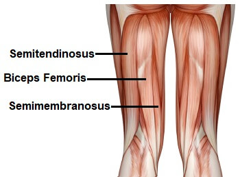
Muscles in the knee diagram. Our interactive 3d knee diagram is an informative 360 degree rotating model.Functional anatomy and clinical relevance of the leg muscles.The hamstring muscles are three muscles at the back of the thigh that affect hip and knee movement.
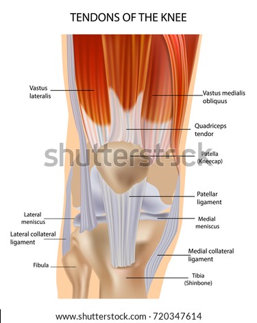
Muscles of the knee to atrophy post operatively responsible for last 10 15o of knee extension vastus medialis obliquus.Each of the 6 sections bones connective tissue 1 connective tissue 2 deep muscles muscles skin can be opened up rotated left or right and viewed more closely.Deep posterior muscles of the leg this article explains the anatomy function and clinical relevance of the deep muscles of the leg.
The knee joins the thigh bone femur to the shin bone tibia.Its complexity and its efficiency is the best example of gods creation.The knee is the meeting point of the femur thigh bone in the upper leg and the tibia shinbone in the.
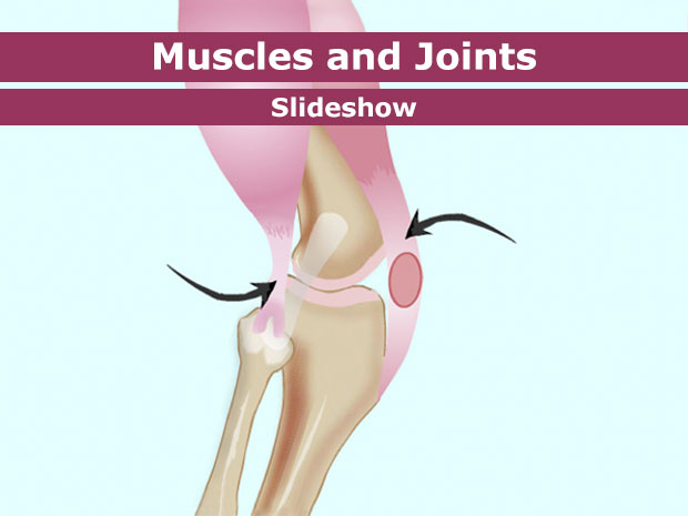
The knee is a complex joint that flexes extends and twists slightly from side to side.Knee joint is one of the most important hinge joints of our body.Our knee muscles are responsible for initiating and controlling movement of the knee and the kneecap.
The middle child of the lower extremity.Myology of the knee.The anatomy of the knee consists of bones muscles nerves cartilages tendons and ligaments.
The muscles go into spasm and even the slightest movements are painful.Tendons usually attach muscle to bone.Structure function of the knee one of the most complex simple structures in the human body.
See diagrams and start learning.Overview of the muscles of the leg and knee.Learn this topic now at kenhub.
They also work work with the various buttock thigh and calf muscles to help control the hip and foot.In the knee the quadriceps and patellar tendon can sometimes tear.They begin under the gluteus maximus behind the hip bone and attach to the tibia at the knee.
X rays can easily confirm the injury and surgery depends on the degree of displacement and type of fracture.

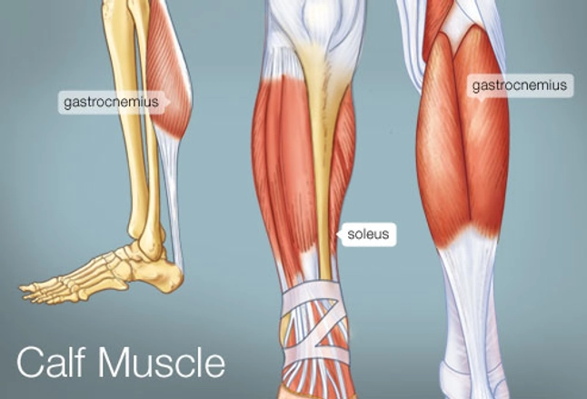
The Calf Muscle Human Anatomy Diagram Function Location


Name The Thigh Muscles Quiz By Jenniferstai13

Acl Solutions Acl Knee Anatomy And Diagram Images

Knee Muscles Anatomy Knee Anatomy Muscles Knee Anatomy The Jullianus

Duke Anatomy Lab 16 Upper Lower Limb Joints

Your Muscles For Kids Kidshealth

Knee Joint Anatomybonescartilagesmusclesligamentstendons Quadriceps

Bent Knee Diagram Wiring Diagram

Knee Muscles Knee Pain Explained

Knee Anatomy Gastrocnemius Muscle Tear Calf Muscles Pathology

Illitibial Band Syndrome Symptoms Treatment Exercises

Knee Anatomy Muscles Tendons Muscle Structure Stock Vector Royalty

Treating Knee Pain And Iliotibial Band Syndrome Itbs

Knee Anatomy Pictures Bones Ligaments Muscles Tendons Function

Sartorius Muscle Attachments And Actions As It Relates To Yoga

Is There Something Wrong With My Kneecap Coastal Orthopedics

Paraspinal Muscles Anatomy Human Muscles Archives Page 31 Of 53

Knee Wikipedia
Experiencing Knee Pain Heres What You Should Know About Patellar
Tendons connect the knee bones to the leg muscles that move.

Muscles in the knee diagram. Our interactive 3d knee diagram is an informative 360 degree rotating model.Functional anatomy and clinical relevance of the leg muscles.The hamstring muscles are three muscles at the back of the thigh that affect hip and knee movement.
Muscles of the knee to atrophy post operatively responsible for last 10 15o of knee extension vastus medialis obliquus.Each of the 6 sections bones connective tissue 1 connective tissue 2 deep muscles muscles skin can be opened up rotated left or right and viewed more closely.Deep posterior muscles of the leg this article explains the anatomy function and clinical relevance of the deep muscles of the leg.
The knee joins the thigh bone femur to the shin bone tibia.Its complexity and its efficiency is the best example of gods creation.The knee is the meeting point of the femur thigh bone in the upper leg and the tibia shinbone in the.
The knee is a complex joint that flexes extends and twists slightly from side to side.Knee joint is one of the most important hinge joints of our body.Our knee muscles are responsible for initiating and controlling movement of the knee and the kneecap.
The middle child of the lower extremity.Myology of the knee.The anatomy of the knee consists of bones muscles nerves cartilages tendons and ligaments.
The muscles go into spasm and even the slightest movements are painful.Tendons usually attach muscle to bone.Structure function of the knee one of the most complex simple structures in the human body.
See diagrams and start learning.Overview of the muscles of the leg and knee.Learn this topic now at kenhub.
They also work work with the various buttock thigh and calf muscles to help control the hip and foot.In the knee the quadriceps and patellar tendon can sometimes tear.They begin under the gluteus maximus behind the hip bone and attach to the tibia at the knee.
X rays can easily confirm the injury and surgery depends on the degree of displacement and type of fracture.

The Calf Muscle Human Anatomy Diagram Function Location

Name The Thigh Muscles Quiz By Jenniferstai13
Acl Solutions Acl Knee Anatomy And Diagram Images
Knee Muscles Anatomy Knee Anatomy Muscles Knee Anatomy The Jullianus
Duke Anatomy Lab 16 Upper Lower Limb Joints

Your Muscles For Kids Kidshealth

Knee Joint Anatomybonescartilagesmusclesligamentstendons Quadriceps

Bent Knee Diagram Wiring Diagram

Knee Muscles Knee Pain Explained

Knee Anatomy Gastrocnemius Muscle Tear Calf Muscles Pathology

Illitibial Band Syndrome Symptoms Treatment Exercises

Knee Anatomy Muscles Tendons Muscle Structure Stock Vector Royalty

Treating Knee Pain And Iliotibial Band Syndrome Itbs

Knee Anatomy Pictures Bones Ligaments Muscles Tendons Function

Sartorius Muscle Attachments And Actions As It Relates To Yoga

Is There Something Wrong With My Kneecap Coastal Orthopedics

Paraspinal Muscles Anatomy Human Muscles Archives Page 31 Of 53

Knee Wikipedia